Google analytics Link

Preferential Preservation of Antiforms During Folding of Biotite-Amphibolite Anatexites, Ladakh, NW India


Roberto Weinberg, Monash University, Australia


[an error occurred while processing this directive]


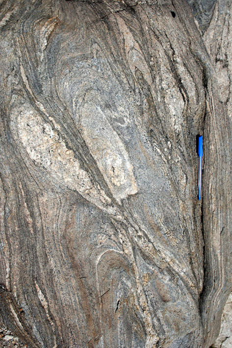
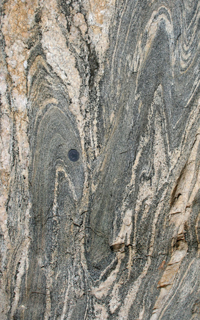
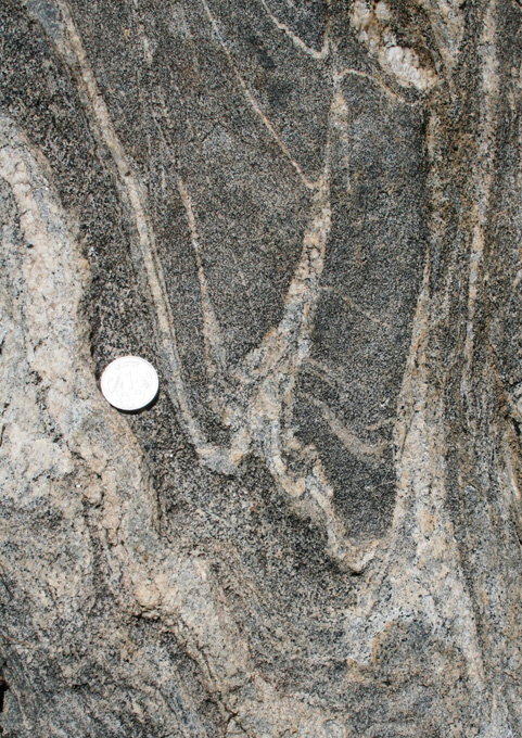
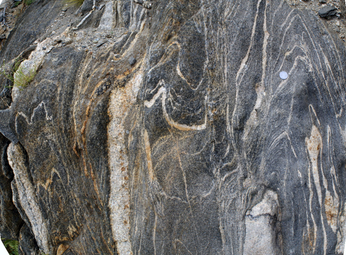
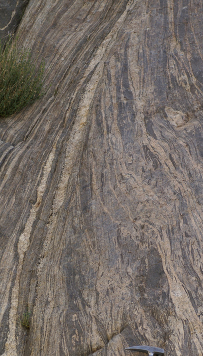
This page focuses on two main aspects of folding during partial melting of biotite amphibolites: a) the preference for preservation of antiforms rather than synforms; b) truncation and continuitiy of layering across fold hinges. All photographs taken down plunge of fold axes over a small area of outcrop in the Tangtse gorge in NW Ladakh, centered around coordinates N34 02' 29.4" E78 13' 18.1".


Note to author: preference for antiform and destruction of synform, 4759 needs to be accompanied by a line drawing, 4779good, IMG_4776zoom, 4784good synform and melting pushing up, stitched_894, DSC00879good, 988, 989, 991, 976, 868, 871


a)  Preferential preservation of antiformal hinges and destruction of synforms


antiform

antiform

Figure 1a. (879 outcrop). Three antiforms separated by narrow axial planar leucosomes occupying the position of synforms. Figure 1b.  (4779 outcrop) A pile up of three antiforms caused by the squeesing of intervening synforms and the wedge extrusion of the leucosome-cored antiform in the top left-hand-side.


antiform

antiform and split synformal hinge


Figure 2a. (4776 outcrop). Two antiforms separated by a disrupted synform which has a left limb transposed by mobile leucocratic material. Figure 2b.(4784 outcrop).  A synformal hinge split by the development of  antiforms. Layer-parallel leucosomes in the synform orm axial planar leucosomes in the antiforms. In the larger antiform to the right,  leucosome truncates the right limb of the synform. Interpretation: folding took place in the presence of melt. The upward movement of melt gave rise to the two antiformal leucosomes that break up the synform..This is the process that leads to the break-up, stretching and transposition of synforms such as seen in Fig. 2a.


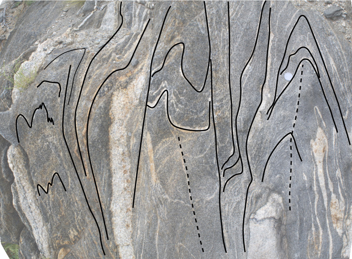
Figure 3a. (Stitched_892-894 outcrop). Three discontinuour sets of folds separated by stretched zones of transposition. The sets on the left and right are antiforms, whereas the central one has both a synform and an antiform. Layer parallel leucosomes have diffuse boundaries with surrounding biotite amphibolite, and diffuse contacts with axial planar leucosomes. Figure 3b. Line drawing. Dashed lines mark narrow axial planar leucosomes, as opposed to the three wider axial planar dykes.


Figure 4a (988 outcrop). Two antiforms separated by a tight, nearly transposed synform. The antiform on the left evolves upwards into an isolated fold hinge zone within tramnsposed surroundings. The antiform on the right continues on to a synform further to the right (not shown). Figure 4b (976 outcrop). Folding of  melano- and leucocratic layers separated by a mesocratic axial planar dyke (left), and a leucocratic axial planar dyke (right). Layering is discontinuous across truncating axial planar dykes and boundaries between dykes and surroundings are diffuse and continuous into layer-parallel leucosomes. The synformal closure on the left-hand-side  and antiformal closure on the right have been disrupted. Interpretation: syn-anatectic folding, where axial planar dykes represent magma escape channels. The mesocratic dyke contains newly grown porphyroblastic hornblende resulting from partial melting of the surrounding biotite-amphibolite. Such hornblende can also be seen in small numbers in the leucocratic dyke, as well as along well-defined layers in the folded rock. See page on amphibolite melting.


Figure 4c (IMG2199 block). Two antiforms.  The intervening synform was used as a slip plane. Figure 4d (3854 ). Figure 4e (3883).


Figure 5 (991 outcrop). This figure shows the less common case of three synforms separated by narrow (right), or  stretched out and transposed (left) antiforms.


PART B: truncation of fold hinge zones


Figure 7  (1332_2005 outcrop). A mesocratic dyke cuts across  a fold.  The dyke has ill-defined layering, compared to the folded rock which has well-defined leuco- and melanocratic layers.


truncated fold

Figure 8a. (871 outcrop). This is a characterisitc feature of many folds in the Tangtse migmatites. There is a break in continuity along the hinge zone of the fold. Truncation of the fold moves from the fold core (lower part of photo) outwards. No leucosome along the axial plane that truncates the fold. Figure 8b.  (873) . Detail of a, showing truncation of layers against the hinge zone in the lower part of the photograph, in contrast to the continuous fold in the upper part.


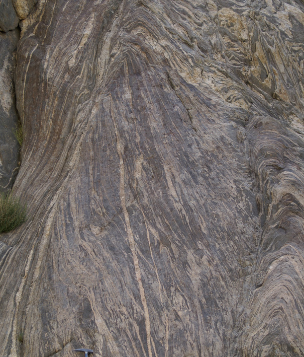
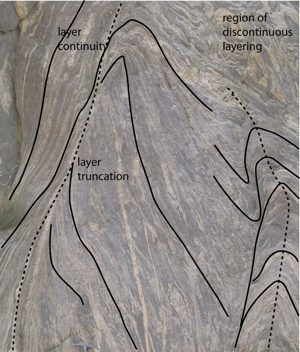
Figure 9a. (897 outcrop). Two large antiforms separated by a narrow synform.  The parasitic antiform on the right is moulded against the large antiform on the left. The core of the large antifrom is truncated by an axial planar leucosome (see detail in Fig. 9c). Hammer for scale at bottom left. Figure 9b. Line drawing. Dashed lines mark hinge line.


truncated fold

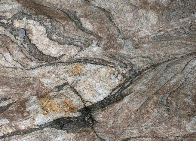
Figure 9c (900 outcrop). Detail of Fig. 9a showing layer truncation against a leucocratic dyke in the hinge zone of the fold. Figure 10b. (868 outcrop). This is a characterisitc feature of many folds in this migmatitie. There is a break in continuity along the hinge zone of the fold. Like here, the core of the fold (lower part is truncated) while the outer hinge zones are continunous across the fold hinge. In this case there is no leucosome in the truncated area of the hinge zone.


truncated fold

truncated fold

Figure 10a (993 outcrop). Truncation along the hinge line of a fold. The left-hand-side has folds with wavelengths of 20cm, which become transposed upwards, while the fold on the right-hand-side is at a much larger (multimetric scale). The two fold systems are disconnected by the truncating leucogranitic dyke in the hinge zone. At a larger scale, not shown on this photograph, the layering become continuous across the fold hinge zone from the limb on the right to the one on the left.  (ADD A LINE SKETCH) Figure 10b (992). Detail of 10a.